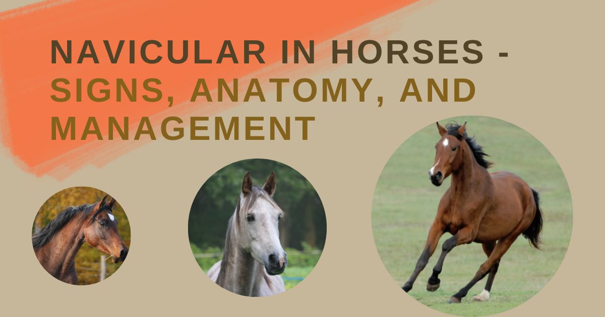
What is Navicular in Horses?
Navicular in horses refers to an umbrella term describing heel pain. The preferred terms for caudal heel lameness are podotrochlosis, podotrochleitis, or navicular syndrome. The podotrochlear apparatus may be affected by several possible causes of pain in and around the navicular bone and its associated structures.
The term may appear simple, but the condition is very complex. Navicular disease in horses can affect the navicular bone, the navicular bursa, and the associated soft tissue structures.
Anatomy of the Equine Navicular Region
The navicular region’s anatomy centers around the navicular bone. The navicular bone is also known as the distal sesamoid bone. The bone’s shape resembles a boat with its small flattened surface across the back of the coffin joint.
The navicular bone is attached to the pedal bone by the short and robust impar ligament and the pastern joint by the suspensory ligaments.
The lower surface of the navicular bone acts as a pulley for the deep digital flexor tendon (DDFT). The navicular bursa acts as a small fluid cushion between the DDFT and the navicular bone.
The Causes of Equine Navicular Disease
Intermittent lameness in horses causes immense frustration for patients and owners alike. The exact cause of navicular in horses has not yet gained a fully supported scientific explanation. According to the pathological changes reported in affected horses, researchers have two possible theories.
The two hypotheses around the causes of the various pathological changes reported in navicular disease include the vascular theory and the biomechanical theory.
- The Vascular Theory: arteriosclerosis and thrombosis of the main nutrient arteries in the distal portion of the navicular bone lead to ischaemic necrosis and bone resorption following reperfusion. This theory is unsupported by histological data, indicating increased bone turnover rather than ischemia and necrosis occurs.
- The Biomechanical Theory: abnormal mechanical forces between the deep digital flexor tendon and the navicular bone’s flexor surface results in cumulative, degenerative changes of the flexor surface and the spongiosa.
Biomechanical data indicates that the force on the navicular bone is increased by contraction of the deep digital flexor tendon in the early phase of the horse’s stride. Histological evidence reveals similar changes to navicular subchondral bone and spongiosa, resulting in osteoarthritis in the low-motion joints.
Noticeable changes include generalized sclerosis and thickening of the subchondral bone plate, focal bone destruction and replacement by granulation tissue, and fibrosis with venous hypertension in the spongiosa.
Navicular is considered a chronic degenerative disease. Lameness occurs due to degeneration affecting the navicular bone’s fibrocartilage, the navicular suspensory ligaments, the impar ligament, the deep digital flexor tendon (DDFT), and the navicular bursa.
The lame horse may be affected in a single limb, but both front hooves are often afflicted. It is rare for the hindquarters to be affected, but veterinarians have documented some cases.
A horse’s distal limb conformation may also play a part in the development of navicular. The relationship between the surrounding navicular structures and hoof angles also places horses with poor hoof care at risk of developing the condition.
Some structures associated with navicular disease in horses include excessively long toes, under-run heels, and a “broken back” hoof-pastern axis.
Any horse can suffer from navicular syndrome, but some breeds that may be predisposed to developing the condition include Thoroughbreds, Quarter Horses, and Warmbloods. Diagnosis of the disease often occurs in mature horses between 4 and 15 years of age.
High-performance horses that undergo excessive strain or sport-related injury from highly physical disciplines can compound the problem with navicular syndrome development. Activities involving hard turns, quick stops, sideway movements, or jumping exacerbate underlying navicular issues.
Signs of Navicular in Horses
When assessing navicular in horses, symptoms may range from unilateral or bilateral low-grade lameness to subtle signs of head bobbing at a trot.
Due to the slow and chronic nature of navicular disease, owners or riders may only notice a subtle, intermittent lameness that increases in frequency when horses are worked on hard ground or asked to turn a small circle.
A short stride characterizes chronic lameness caused by navicular with toe-landing and pain from the center third of the frog.
Diagnosing Navicular in Horses
Veterinarians use a combination of a patient’s clinical history and symptoms, a complete physical examination, nerve blocks, and diagnostic imaging such as radiographs or an MRI to diagnose navicular disease.
Vets examine the hoof visually and determine any abnormalities through palpation and flexion tests. The hoof biomechanics assessment includes the wall, frog, and foot shape.
Hoof testers are an essential tool to help with the diagnosis because some horses will be sensitive over the frog and heels if they have navicular pathology.
Physical exams for a lameness work-up require the horse to be lunged or under saddle to evaluate the horse’s gait. Horses should be examined within an arena or any other soft surface and on a hard surface to properly assess any differences in the horse’s gait.
Often a horse develops a head bob and increased lameness in the affected leg on a hard surface because of the increase in concussive forces.
Flexion tests of the distal limb will flex positive in chronic foot lamenesses. Using a palmar digital nerve block helps gauge lameness in bilaterally affected forelimbs. This test also helps owners realize the severity of the lameness through a visual comparison between the blocked and lame limbs.
A wedge test uses a block to elevate the frog, toe, and hoof walls, the horse then trots a short distance, and the lameness is evaluated and rated after each elevation change. This test helps to localize the horse’s perceived pain.
Veterinarians use nerve blocks to localize navicular disease if the block alleviates lameness in the DIP joint (coffin joint,) back half of the foot or the navicular bursa.
Diagnostic imaging plays a vital role in chronic lameness workups because it allows the clinician to visualize the joints, the bones, and the soft tissue supportive structures through radiographs or magnetic resonance imaging (MRI).
MRIs are the golden standard test, but they are a very costly procedure. Changes seen in navicular disease in horses include the following:
- ‘Classical’ navicular disease: degenerative changes of the navicular bone’s palmar fibrocartilage and flexor cortex. The bursal surface of the superficial, deep digital flexor tendon and navicular bursa also show degeneration and inflammatory changes.
- Distal border fragments and navicular bone remodeling: bone fragments occur at the lateral or medial angles of the distal bone border. Bone remodeling and potential fragmentation can result in lameness.
- Medullary bone necrosis: sometimes, primary medullary injury occurs, and notable medullary adipose tissue edema and necrosis.
- Desmitis or enthesopathy of the navicular suspensory ligaments or impar ligament. These changes are less common than bone-related abnormalities.
Radiographs are sometimes more affordable and help to reveal boney changes in the navicular structures.
Making a radiological diagnosis requires good quality radiographs and several views to interpret the navicular bone changes or abnormalities correctly. The foot requires proper preparation, correct positioning, and accurate settings for optimal execution of the x-ray in the field or a hospital.
Veterinarians must heed caution when interpreting the x-rays to avoid creating radiological artifacts. Possible abnormalities include the following:
- Navicular bone proximedial and prolateral enthesiophytes.
- Distal or proximal extension of the flexor border of the bone.
- Bone fragments at the distal edge of the bone.
- Variably shaped radiolucent zones at the bone’s distal edge, eight or more zones indicate pathology.
- Subtle radiolucent areas in the bone’s spongiosa may or may not communicate with the deep digital flexor tendon cortex.
- Novel bone formation at the sagittal ridge.
- Increases in flexor cortex thickness.
- Spongiosa sclerosis.
- Presence of a bipartite bone.
Veterinarians must interpret the radiographic changes with the horse’s breed, age, and workload born in mind. Some radiographic changes may be within normal limits or significantly altered depending on the criteria above.
Vets use bursograms to differentiate between normal tendon adhesions and possible tendon injuries if the injected dye communicates with the bursa. Considerations for iatrogenic injury to the deep digital flexor tendon are noteworthy, even though the occurrence is rare.
Ultrasound examinations of the supportive soft tissue structures such as the deep digital flexor tendon and impar ligament are also helpful in visualizing the possible origin of the lameness. Ultrasonography is cost-effective and non-invasive, but it requires a skilled operator to diagnose tendon pathology accurately.
Managing Navicular Disease in Equines
Veterinarians can manage navicular disease with different treatments, but no treatments are curative. A multimodal approach to managing the symptoms and preventing flare-ups is vital.
Resting lame horses is the first step to alleviating symptoms. Once a horse becomes noticeably lame, the best option to avoid exacerbating symptoms or secondary complications is to stop training until a vet has seen them.
In painful patients, veterinarians prescribe non-steroidal anti-inflammatory drugs (NSAID) such as phenylbutazone or firocoxib to alleviate discomfort. Horses with underlying gastric ulcers or possible renal issues should be given conservative NSAID doses and monitored closely for adverse side effects.
The use of intra-articular corticosteroid injections shows improvement in a third of horses for an average of 2 months. Injections into the navicular bursa can resolve lameness for up to 4 months, but there are risks associated with intrabursal injections, such as deep digital flexor tendon rupture.
Chronic treatment with isoxsuprine and aspirin improved blood supply to the navicular bone. Unfortunately, isoxsuprine has little therapeutic value when administered orally. Warfarin used in the past predisposes horses to bleeding tendencies.
Shoeing and trimming show the most promise when it comes to podotrochlosis management. Veterinarians must consult with a farrier to synergistically combine radiographic findings and guide trimming and shoeing decisions.
A horse’s foot shape and conformation need individual assessments to determine the best fit to alleviate symptoms and avoid flare-ups. There is no set protocol, so treatments must find a basis according to the hoof’s biomechanical needs. The main aim is to relieve pain and maintain functionality while considering disease severity and the horse’s working discipline.
An excellent example of a shoe suited to a horse with navicular syndrome is a wedged shoe with a welded heel plate. This shoe uses the navicular bone as a fulcrum and decreases the tension on the deep digital flexor tendon.
The shock transmitted through the frog from concussive forces becomes blocked by the heel plate, thus preventing the force from reaching the navicular area.
Surgical management of navicular in horses ranges from bursoscopy and arthroscopy to neurectomy procedures. Each case depends on the cause of the horse’s symptoms.
A palmar digital neurectomy desensitizes the nerves of the heel and acts as a semi-permanent pain relief option. Surgery of this nature is a last resort as post-operative complications occur frequently and include deep digital flexor tendon rupture or painful neuroma formations.
A neurectomy is a good solution for horses intended for low-level, nonstrenuous work. The procedure prolongs their ability to be used as working horses while maintaining an acceptable level of comfort.
Bisphosphonates are drugs the FDA recently approved as an additional option when treating navicular.
Biphosphonates act by reducing bone remodeling and blocking excessive bone resorption. This medication is only effective in horses with bone-related navicular symptoms. The drug can cause colic, may negatively affect renal function, and should not be administered concomitantly with NSAIDs.
Extracorporeal shock wave and laser therapy also show promising results in pain management for horses with soft tissue injuries. The shock waves reduce healing time in ligament and tendon injuries.
If a horse’s symptoms become manageable and the lameness abates, getting a horse back into work is essential. Trainers and riders must restore hoof biomechanics through constructive work and everyday stresses on appropriate surfaces.
Clinicians cannot cure navicular disease, but owners can manage the condition if they comply with regular farrier visits and veterinary follow-ups.
Can Navicular in Horses be Prevented?
Undiagnosed podotrochlosis leads to chronic lameness, which results in permanent changes to the heel conformation. Preventing the progression of lameness is vital in avoiding chronic lameness. Horse owners need to seek out the services of a skilled and reliable farrier to provide a regular shoeing or trimming schedule that works well with the horse’s attending vet.
As soon as a rider or trainer notices a horse’s change in attitude to work or an altered gait, it is vital to take action and consult with a vet. Prevention starts with early detection.
Foot balance and heel support are the cornerstones of lameness prevention. Unfortunately, due to the nature of navicular syndrome, it is not always possible to entirely prevent the symptoms as the causes remain undefined.
Any foot shape in a horse can develop the disease. A horse’s natural foot angle allows the correct bone alignment. Farriers should trim hooves with a short toe to let the foot break over easily.
Nutrition is key to maintaining a horse’s body condition and keeping hooves healthy. An overweight horse places additional strain on joints, especially if they are predisposed to navicular disease. Navicular horse supplements aim to support joint health and are a great addition to a horse’s diet to help decrease or manage inflammation and protect joints.
Detecting navicular in a young horse has a better prognosis if they receive treatment early and may even maintain their performance abilities. Older horses have a more guarded prognosis as they are most likely in the later stages of the disease.
Can a Horse With Navicular Jump?
Navicular is a chronic degenerative condition, so even with good pain management and corrective shoeing – intermittent lameness is a long-term reality for an affected horse. Chronic lameness may lead to frustration for trainers and riders trying to condition a competing horse.
If the horse is coping with conservative management of the condition, then here are a few tips to try and avoid flare-ups:
- Corrective trimming every six weeks.
- Shoeing needs to encourage an early break-over and provide good heel support.
- Avoid training on rocky or uneven ground and dismount before any steep hills.
- Maintain a healthy weight as an overweight horse places more strain on their joints.
- Try to avoid harsh surfaces when considering a competition arena.
Conclusion
Navicular disease is degenerative, and treatments mainly focus on delaying degeneration and managing pain and lameness. Some horses may never return to work, while others continue to compete and only stop when their symptoms progress into the chronic phase of the condition.
Podotrochlosis is a complex condition to manage. Owners cannot approach treatments with a one-size-fits-all attitude. The intermittent bouts of lameness and frequent vet and farrier visits can become quite costly, and it becomes a big financial responsibility to comply with the intensive needs of a horse with navicular disease demands.
A collaborative effort is required between owners, trainers, riders, vets, and farriers to gain successful results and avert lameness episodes.
Unfortunately, it is difficult to prevent due to the unknown etiology of navicular disease in horses. Early lameness detection, good conditioning, regular follow-ups, and frequent farrier visits are the best way to keep navicular at bay.